Microtia and Atresia
Microtia is a congenital disorder that causes a deformity in which the outer ear (called “pinna”) is underdeveloped or absent. It can affect one or both ears. The word microtia translates literally to mean “small ear”, and young children and their families will often call the affected ear a “small ear”. A fully formed ear or a reconstructed ear is often called a “big ear”.
Atresia is the absence of an external ear canal, and in many cases the ear drum and ear bones can also be affected. This causes a conductive hearing loss because there is no canal through which sound can travel.
Microtia is often, but not always, accompanied by atresia because a baby’s outer ear and ear canal develop together during pregnancy. In some cases the microtia may be part of a craniofacial condition, meaning that the child may also have other head or face structure deformities. In these cases the microtia is best assessed as part of the whole craniofacial condition.
Just as every ear is different, every microtia ear remnant is also different. The wonderful news is that microtia treatments are evolving quickly and it is now possible for a patient to receive an ear implant that is totally customised to their head shape.
Dr Dusseldorp performed the first custom 3D printed ear implant (SU-POR®) procedure in Australia in March 2019. This first surgery was for a 4 year old boy, Maxim. You can read Maxim’s story and watch the Channel 10 News coverage of his journey.

Since then Dr Dusseldorp has performed similar surgeries for children aged at least 4 years old, and he is also the first to combine the custom 3D ear implant procedure with a bone conduction hearing aid implant to treat atresia. This innovative approach means that a child is only exposed to one operation and can immediately start their recovery period after just one night in hospital.
Microtia Treatment Options
There are a range of treatments for microtia and each is explained below. The custom 3D printed implant is the most recent innovation, and it is the treatment option requiring least surgery and recovery time, with less pain. It also offers the most aesthetically refined outcome and can be performed at a younger age than some other treatment options.
Cartilage Framework
This method can be used for a range of different ear reconstruction needs, including microtia and ears affected by some form of trauma. The method utilises a section of a patient’s rib cartilage, which is harvested during their first operation, sculpted by the surgeon into a shape that most suits the patient’s need, and put in place under the skin. During a second operation 6 weeks later the ear is adjusted to take on a 3 dimensional shape. This method can only be used for a child who has reached at least 8 years of age, and requires 4 days in hospital for recovery after the first operation.
Note that microtia surgery is not just an option for children. One of Dr Dusseldorp’s first patients was an adult in the USA who elected to treat his microtia because of the improved outcomes of more recent techniques. Read more about Shawn’s experience. You can also see more examples of the cartilage framework option from Dr Dusseldorp’s mission in Vietnam in March 2019.

MEDPOR®
MEDPOR® stands for “microporous high-density polyethylene implant”. It is the first evolution of 3D printed implants and is used for a wide array of anatomical reconstruction needs, not just ears. This porous polyethylene implant is bio-compatible with a patient’s tissue, and during surgery the patient’s own skin is grafted over the implant. This procedure has been offered for 20 years in the USA and can be performed on children upward of about 4 years of age. The full procedure can be performed in just one surgery and it is less invasive and less painful than the Cartilage Framework method. MEDPOR® implants are only available in three different sizes.
SU-POR®
SU-POR® is also a bio-compatible porous polyethylene material from which a range of anatomical implants can be created. The difference with SU-POR® is that an implant can be custom 3D printed, so an ear shape is not restricted to the 3 sizes available in MEDPOR®.
Dr Dusseldorp believes there are several important benefits of the 3D custom printed ear reconstruction technique. These include a more natural aesthetic appearance, a less invasive approach, fewer surgeries performed at a younger age than the Cartilage Framework method, and less physical and psychological pain. Importantly, it is also possible to combine BAHA hearing device implantation simultaneously with microtia repair, reducing the overall number of surgeries required to treat both microtia and atresia.
Custom 3D printed ear reconstruction was launched in Australia in March when Dr Dusseldorp performed the first case of unilateral microtia in the context of hemi-facial microsomia in a four year old child. Prior to this, similar procedures have been performed over 1500 times in the United States, which is where Dr Dusseldorp trained and has operated previously.
The magic of 3 dimensional technology is not reserved only for the printing of the ear implant. 3D scanning is used to create a comprehensive model that can be used by surgeons to design a series of ear models to best match a patient’s full head shape. You can see the 3D scan that was done for Maxim, and you can even interact with it yourself!
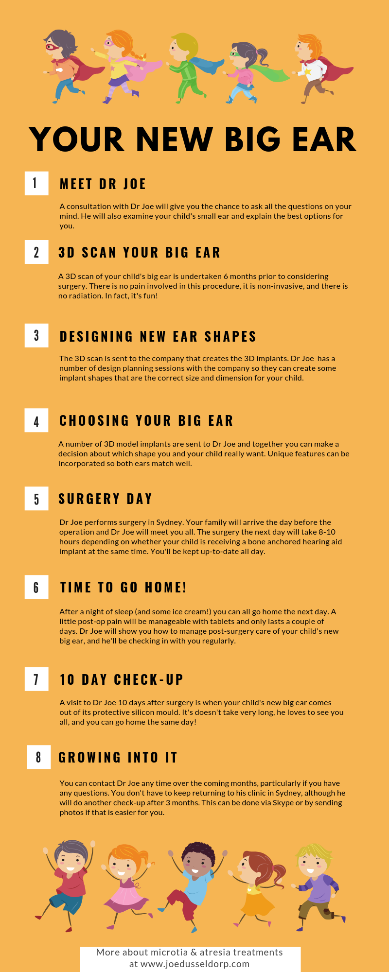
More about the custom 3D printed ear implant process
The infographic below provides some details on the steps your child and your family will go through.